Showing posts with label pediatric blog. Show all posts

Thursday, March 15, 2012

HbA1c and Estimated Average Glucose (eAG)

Source of this information: ADA and http://www.ngsp.org/A1ceAG.asp


There is a very predictable relationship between HbA1c and AG. 
Understanding this relationship can help patients with diabetes and their health-care providers set day-to-day targets for AG based on HbA1c goals
Fasting glucose should be used with caution as a surrogate measure of AG. 
Finally, it is important to remember that HbA1c is a weighted average of glucose levels during the preceding 4 months. Unless the patient’s glucose levels are very stable month after month, quarterly measurement is needed to insure that a patient's glycemic control remains within the target range. 

How does HbA1c relate to average glucose (AG)?

In the Diabetes Control and Complications Trial or DCCT (New Engl J Med 1993;329:977-986) study of patients with Type 1 diabetes, quarterly HbA1c determinations were the principal measure of glycemic control; study subjects also performed quarterly 24-hour, 7-point capillary-blood glucose profiles. 
Blood specimens were obtained by subjects in the home setting, pre-meal, 90 minutes post-meal, and at bed-time. In an analysis of the DCCT glucose profile data (Diabetes Care 25:275-278, 2002), mean HbA1c and AG were calculated for each study subject (n= 1439). Results showed a linear relationship between HbA1c and AG 
(AG(mg/dL) = ( 35.6 x HbA1c ) - 77.3), with a Pearson correlation coefficient (r) of 0.82.
CALCULATOR: The following link will help to do the Estimated Average Glucose Calculation
http://professional.diabetes.org/GlucoseCalculator.aspx :


HbA1c (%)
eAG (mg/dL)
eAG (mmol/l)
5
97
5.4
6
126
7.0
7
154
8.6
8
183
10.2
9
212
11.8
10
240
13.4
11
269
14.9
12
298
16.5

Tuesday, March 13, 2012

History of HbA1c and what does it do?

History:
Hemoglobin A1c was first separated from other forms of hemoglobin by Huisman and Meyering in 1958.  It was first characterized as a glycoprotein by Bookchin and Gallop in 1968. Its increase in diabetes was first described in 1969 by Samuel Rahbar The use of hemoglobin A1c for monitoring the degree of control of glucose metabolism in diabetic patients was proposed in 1976 by Anthony Cerami, Ronald Koenig and coworkers.
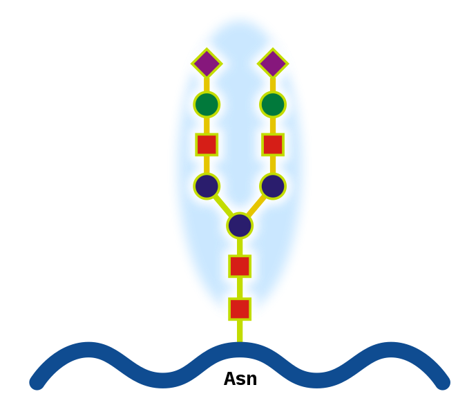
N-linked protein glycosylation (N-glycosylation of N-glycans) at Asn residues (Asn-x-Ser/Thr motifs) in glycoproteins.


What are Glycoproteins? 
They are proteins that contain oligosaccharide chains (glycans) covalently attached to polypeptide side-chains. The carbohydrate is attached to the protein in a cotranslational orposttranslational modification. This process is known as glycosylation. In proteins that have segments extending extracellularly, the extracellular segments are often glycosylated. Glycoproteins are often important integral membrane proteins, where they play a role in cell–cell interactions. Glycoproteins are also formed in the cytosol, but their functions and the pathways producing these modifications in this compartment are less well understood


Why do we measure HbA1c every 3 months?

In the normal 120-day lifespan of the red blood cell, glucose molecules react with hemoglobin, forming glycosolated hemoglobin. In individuals with poorly controlled diabetes, the quantities of these glycosolated hemoglobins are much higher than in healthy people.
Once a hemoglobin molecule is glycosolated, it remains that way. A buildup of glycosolated hemoglobin within the red cell, therefore, reflects the average level of glucose to which the cell has been exposed during its life-cycle. Measuring glycosolated hemoglobin assesses the effectiveness of therapy by monitoring long-term serum glucose regulation. The HbA1c level is proportional to average blood glucose concentration over the previous four weeks to three months. Some researchers state that the major proportion of its value is related to a rather shorter period of two to four weeks.
Image source: http://adiscar-adiscar.blogspot.com

We will talk about measuring HbA1c tomorrow

Monday, March 12, 2012

Tummy Tuck and Stretch Marks!

Medical Term for Strech Marks: Striae
Stretch marks or striae (singular stria), are a form of scarring on the skin with an off-color hue. They are caused by tearing of the dermis, which over time may diminish, but will not disappear completely.
Stretch marks are often the result of the rapid stretching of the skin associated with rapid growth.
Stretch marks can appear anywhere on the body, but are most likely to appear in places where larger amounts of fat are stored. Most common places are the abdomenbreasts, upper arms, underarms, backthighs , hips, and buttocks. They pose no health risk in and of themselves.


In CUSHING SYNDROME, the glucocorticoid hormones responsible for the development of stretch marks affect the dermis by preventing the fibroblasts from forming collagen and elastin fibers, necessary to keep rapidly growing skin taut. This creates a lack of supportive material, as the skin is stretched and leads to dermal and epidermal tearing.
A new modality, fractional laser resurfacing, offers a novel approach to treating striae. Using scattered pulses of light only a fraction of the scar is zapped by the laser over the course of several treatments. This creates microscopic wounds. The body responds to each treatment by producing new collagen and epithelium


Picture courtesy: Derm Atlas


Abdominoplasty or "tummy tuck" is a cosmetic surgery procedure used to make the abdomen more firm. The surgery involves the removal of excess skin and fat from the middle and lower abdomen in order to tighten the muscle and fascia of the abdominal wall. 

Sunday, March 11, 2012

Epistaxes ( Nose bleeds) - Anterior Nose bleed

This is probably one of the scariest medical problem a parent or even an adult experiences and many a times it is innocuous.
Cause is local or generalized (systemic)
Broadly divided to Anterior and Posterior nose bleeds.
Today the discussion will be anterior nose bleeds


Nosebleeds begin in the lower part of the septum, (wall that separates the two nostrils ). The septum has tiny blood vessels that can break by nose blowing or nose picking. Nosebleeds coming from the front of the nose,  (anterior nosebleeds) often begin with blood out one nostril usually when sitting or standing.

Anterior nosebleeds are common during dry climates or during the winter when dry, heated indoor air dehydrates the nasal membrane. Dryness result in crusting, cracking, and finally bleeding. 
This bleeding can be prevented by placing a slight application of petroleum jelly or an antibiotic ointment on the end of a fingertip and then rubbing it inside the nose, especially on the septum.

ADVISE TO STOP BLEEDING
Stay calm, or help the child (or the adult) to calm down. Agitation makes bleeding more profuse.
Keep head higher than the level of the heart. Sit up.
Leaning slightly forward helps the blood not going into the throat
Gently blow any clotted blood out of the nose.  N
asal decongestant spray can help.
Using the thumb and index finger, pinch all the soft parts of the nose.  
Do not pack the inside of the nose with gauze or cotton.
Hold the position for five minutes. 
If bleeding persists, hold an additional 10 minutes.
Here is the first aid demo: from YOUTUBE
Surgical procedure for severe nose bleeding

Saturday, March 10, 2012

Respiratory Syncytial Virus - How to prevent my severity?


How can I help prevent the spread of RSV?

RSV is spread by coming in close contact with an infected person and the droplets produced when the person coughs or sneezes.  Careful hand washing with soap and warm water is the best way to prevent the spread of RSV. 

Palivizumab (brand name Synagis) is a monoclonal antibody produced by recombinant DNA technology. It is used in the prevention of respiratory syncytial virus (RSV) infections. It is recommended for infants that are high-risk because of prematurity or other medical problems such as congenital heart disease.
Palivizumab is a humanized monoclonal antibody (IgG) directed against an epitope in the A antigenic site of the F protein of RSV. Palivizumab reduced the risk of hospitalization due to RSV infection by 55%. Palivizumab is dosed once a month via intramuscular (IM) injection, to be administered throughout the duration of the RSV season.
Palivizumab targets the fusion protein of RSV, inhibiting its entry into the cell and thereby preventing infection.

The American Academy of Pediatrics (AAP) has published recommendations for the use of palivizumabUpdated AAP recommendations were published in 2009. Palivizumab  is used only for prevention, not for treatment, and once initiated for a given RSV season (usually November–March), it should be continued for the full duration of that season.
Reasons to consider palivizumab prophylaxis include:
Prematurity
  • ≤ 28 weeks gestation and < 12 months of age at the start of RSV season
  • 29-32 weeks gestation and < 6 months of age at the start of RSV season
  • 32-35 weeks gestation and < 3 months of age at the start of RSV season, if there is a risk factor (child care attendance or sibling younger than 5 years old)
Chronic lung disease of prematurity
  • Chronic lung disease still requiring oxygen/medication, for the first and second RSV seasons
  • Chronic lung disease that required oxygen/medication within the 6 months preceding RSV season, for the first RSV season
Congenital heart disease
  • Cyanotic heart disease, for the first 24 months of life
  • Moderate to severe pulmonary hypertension, for the first 24 months of life
  • Congestive heart failure requiring medication, for the first 24 months of life
  • Children who have undergone open heart surgery during RSV season, for one additional dose after cardiopulmonary bypass (only if they still meet one of the other criteria)
Other conditions where prophylaxis might be considered but inadequate data is available:
  • Immunocompromise
  • Cystic fibrosis
Of note, a course of palivizumab is quite expensive, and the above recommendations were written based on estimates of its overall cost-effectiveness for preventing severe RSV disease.


Thursday, March 8, 2012

Who am I and why should you be scared of me now?

I am a very common virus that leads to mild, cold-like symptoms in adults and older healthy children. But I am bad with young babies. Most infants have been infected by me before their second birthday.
I love the season from October to Spring (my season!)

More severe  disease with me may occur in the following infants:
  • Premature infants
  • Infants with chronic lung disease
  • Immunodeficient infants
  • Infants with heart disease


The following increase the risk for being affected by me:
  • Day care
  • Tobacco smoke
  • Having school-aged brothers or sisters
  • Crowded conditions

    You get to know about me 4 - 6 days after coming in contact with me.
    Antibiotics cannot make me go away
    If am nice (mild) I go away without treatment.
    Infants and children with a severe infection with me will be in the intensive care unit. Treatment will include:
    • Oxygen ( may need a ventilator)
    • humidified air
    • IV fluids
    I AM Respiratory Syncytial Virus
    Belonging to the Paramyxoviridae, I am virus in an enveloped, spherical, negative-strand RNA virus measuring 120-300 nm

    Diagnosis: You can find me by a direct immunofluorescence test for RSV antigen , and is reported to be 90% sensitive as compared to culture. RSV antigen may appear in the cytoplasm of cells within 8 hours of infection. Viral culture is performed in conjunction with the antigen test. The only acceptable specimen to detect me is a nasopharyngeal aspirate collected in viral transport media.
    Tomorrow I will tell you how to avoid me!!

What is in an egg?

Courtesy: http://dailyfitnessmagz.com


Facts:
1. Chicken egg has less cholesterol than the other eggs!
2. If you remove the egg yolk, you completely remove all the cholesterol. Egg white has NO cholesterol
3. Only animal food has cholesterol
4. One egg has only 75 calories but 7 grams of high-quality protein, 5 grams of fat, and 1.6 grams of saturated fat, along with iron, vitamins, minerals, and carotenoids.
5. The egg has disease-fighting nutrients like lutein and zeaxanthin. These carotenoids may reduce the risk of age-related macular degeneration, the leading cause of blindness in older adults. And brain development and memory may be enhanced by the choline content of eggs.
6. "Designer" eggs may come from chickens that are allowed to roam freely (free range) or whose feed is supplemented with omega-3 fatty acids. Hens given feed that is free of animal products produce vegetarian eggs, while those given all-organic feed produce organic eggs.
7.  Some chicken feed is enriched with canola oil, bran, kelp, flaxseed, marine algae, fish oil, or vitamin E to increase the eggs' healthy omega-3 fatty acid content. Certain types of feed are designed to reduce the saturated and total fat content of the egg yolk. Marigold extract has been used to increase the lutein content of eggs

Wednesday, March 7, 2012

Turner Syndrome: Aortic Coarctation - see the 3D view

Turner's Syndrome: Chromosomal disorder - Turner syndrome affects approximately 1 out of every 2,500 female live births worldwide. It embraces a broad spectrum of features, from major heart defects to minor cosmetic issues. The commenest heart condition is BICUSPID AORTIC VALVE! not Coarctation of Aorta
See this study below:
Pediatr Cardiol. 1999 Mar-Apr;20(2):108-12.: Study shows:
Partial anomalous pulmonary venous drainage (PAPVD; 2.9%), 
Aortic valve disease (stenosis and/or incompetence) (AoVD; 5. 1%), 
Aortic coarctation (AoCo; 4.4%), 
Bicuspid aortic valve (BicAo; 14.7%) are much more frequent in Turner's syndrome than in the normal population, with the difference being statistically highly significant.


High Blood pressure is common because of heart or kidney issues
See and learn below: Magnetic resonance angiography  showing the aortic coarctation.


MR Angiography (MRA) - courtesy -http://turners.nichd.nih.gov/angiography.html

Monday, March 5, 2012

Sleeping positions - today - Tomorrow I will write about the personality traits associated with this


In Professor Chris Idzikowski's survey of 1000 people, he identified six positions and claimed to detect personality traits based on them:
  • Fetus (41%) – curling up in a fetal position. This was the most common position, and is especially popular with women.
  • Log (15%) – lying on one's side with the arms down the side.
  • Yearner (13%) – sleeping on one's side with the arms in front.
  • Soldier (8%) – on one's back with the arms pinned to the sides.
  • Freefall (7%) – on one's front with the arms around the pillow and the head tilted to one side.
  • Starfish (5%) – on one's back with the arms around the pillow
  • Rest of them did not know how they slept

This is a study of 1000 people ( source Wikipedia)

Sunday, March 4, 2012

Pterygium colli deformity (well what I meant was - Webbed Neck!)

Congenital skin fold that runs along the sides of the neck down to the shoulders
This is seen in Turner syndrome and Noonan syndrome, and  Klippel-Feil syndrome
In the newborn period, webbed neck is the loose folds of skin on the neck. 
As the child grows, the skin may stretch out to look like there is either a very short neck or no neck at all.
A 12-year-old female with Noonan syndrome exhibiting a typical webbed neck.
Picture courtesy: Wiki commons

Friday, March 2, 2012

GO away bad cholesterol - Why do we not want LDL?

Lipoproteins: Lipid (fat) + Protein, whose function is to transport lipids (fats) (such as triacylglycerol) around the body in the blood.
There are 5 major groups 


Lipoprotein structure (chylomicron)
ApoA, ApoB, ApoC, ApoE (apolipoproteins); T(triacylglycerol); C (cholesterol); green (phospholipids)
Picture - Wikipedia


Their size varies - small to Big  (chylomicronsVLDLIDLLDL, & HDL)


Low-density lipoprotein (LDL)  enable transport of multiple different fat molecules, including cholesterol, within the water around cells and within the water-based bloodstream


Higher levels of BAD cholesterol AKA type-B LDL particles (as opposed to type-A LDL particles) increase health problems and cardiovascular disease


Blood tests typically report LDL-C - This is a calculated measurement
Direct LDL-C measurement using Nuclear magnetic resonance spectroscopy is superior and more accurate


LDL subtype patterns
LDL particles vary in size and density. Pattern that has more small dense LDL particles, called Pattern B, equates to a higher risk factor for coronary heart disease (CHD) than does a pattern with more of the larger and less dense LDL particles (Pattern A). This is because the smaller particles are more easily able to penetrate the endothelium
Pattern I, for intermediate, indicates that most LDL particles are very close in size to the normal gaps in tthe endothelium.
There is also relation ship between higher triglyceride levels and higher levels of smaller, denser LDL particles and alternately lower triglyceride levels and higher levels of the larger, less dense LDL