Hepatitis B serologic testing involves measurement of several hepatitis B
virus (HBV)-specifi c antigens and antibodies. Different serologic “markers”
or combinations of markers are used to identify different phases of HBV
infection and to determine whether a patient has acute or chronic HBV
infection, is immune to HBV as a result of prior infection or vaccination, or
is susceptible to infection.
Hepatitis B surface antigen (HBsAg):
A protein on the surface of hepatitis B virus; it can be detected in high levels in serum during acute or chronic hepatitis B virus infection. The presence of HBsAg indicates that the person is infectious. The body normally produces antibodies to HBsAg as part of the normal immune response to infection. HBsAg is the antigen used to make hepatitis B vaccine.
Hepatitis B surface antibody (anti-HBs):
The presence of anti-HBs is generally interpreted as indicating recovery and immunity from hepatitis B virus infection. Anti-HBs also develops in a person who has been successfully vaccinated against hepatitis B
Total hepatitis B core antibody (anti-HBc):
Appears at the onset of symptoms in acute hepatitis B and persists for life. The presence of
anti-HBc indicates previous or ongoing infection with hepatitis B virus in an undefined time frame
IgM antibody to hepatitis B core antigen (IgM anti-HBc):
Positivity indicates recent infection with hepatitis B virus (<6 mos). Its presence indicates acute infection.
Tests Results Interpretation
Susceptible
HBsAg negative
anti-HBc negative
anti-HBs negative
Immune due to natural infection
HBsAg negative
anti-HBc positive
anti-HBs positive
Immune due to hepatitis B vaccination
HBsAg negative
anti-HBc negative
anti-HBs positive
Acutely infected
HBsAg positive
anti-HBc positive
IgM anti-HBc positive
anti-HBs negative
Chronically infected
HBsAg positive
anti-HBc positive
IgM anti-HBc negative
anti-HBs negative
Interpretation unclear; four possibilities:
1. Resolved infection (most common)
2. False-positive anti-HBc, thus susceptible
3. “Low level” chronic infection
4. Resolving acute infection
anti-HBs negative
HBsAg negative
anti-HBc positive
Adapted from: A Comprehensive Immunization Strategy to Eliminate Transmission of Hepatitis B
Virus Infection in the United States: Recommendations of the Advisory Committee on Immunization
Practices. Part I: Immunization of Infants, Children, and Adolescents. MMWR 2005;54(No. RR-16
Courtesy: http://www.cdc.gov/hepatitis/HBV/PDFs/SerologicChartv8.pdf

SmartMD (SACKIDMD) is 2 minutes of health update Follow @ Facebook.com/sackidMD or Twitter @ SackidMD. Resource from Dr. Prakasam's SACKID group.
Showing posts with label SmartMD. Show all posts
Showing posts with label SmartMD. Show all posts
Friday, February 10, 2012

Thursday, February 9, 2012
Interesting Appendix!
One of the most common causes of severe acute abdominal pain worldwide.
A correctly diagnosed non-acute form of appendicitis is known as "rumbling appendicitis".
The term "pseudoappendicitis" is used to describe a condition mimicking appendicitis. It can be associated with Yersinia enterocolitica.

Picture shows an acutely inflamed and enlarged appendix, sliced lengthwise.
Rovsing's sign
Continuous deep palpation starting from the left iliac fossa upwards (counterclockwise along the colon) may cause pain in the right iliac fossa, by pushing bowel contents towards the ileocaecal valve and thus increasing pressure around the appendix.
Psoas sign or "Obraztsova's sign"
is right lower-quadrant pain that is produced with either the passive extension of the patient's right hip (patient lying on left side, with knee in flexion) or by the patient's active flexion of the right hip while supine. The pain elicited is due to inflammation of the peritoneum overlying the iliopsoas muscles and inflammation of the psoas muscles themselves. Straightening out the leg causes pain because it stretches these muscles, while flexing the hip activates the iliopsoas and therefore also causes pain.
Obturator sign
If an inflamed appendix is in contact with the obturator internus, spasm of the muscle can be demonstrated by flexing and internal rotation of the hip. This maneuver will cause pain in the hypogastrium.
Dunphy's sign
Increased pain in the right lower quadrant with coughing
Kocher's (Kosher's) sign
From the history given, the appearance of pain in the epigastric region or around the stomach at the beginning of disease with a subsequent shift to the right iliac region.
Differential diagnosis
In children
Gastroenteritis, mesenteric adenitis, Meckel's diverticulitis, intussusception, Henoch-Schönlein purpura, lobar pneumonia, urinary tract infection, new-onset Crohn's disease or ulcerative colitis, pancreatitis, abdominal trauma from child abuse, distal intestinal obstruction syndrome in children with cystic fibrosis, typhlitis in children with leukemia;
Courtesy: Wikipedia: http://en.wikipedia.org/wiki/Appendicitis
A correctly diagnosed non-acute form of appendicitis is known as "rumbling appendicitis".
The term "pseudoappendicitis" is used to describe a condition mimicking appendicitis. It can be associated with Yersinia enterocolitica.
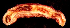
Picture shows an acutely inflamed and enlarged appendix, sliced lengthwise.
Rovsing's sign
Continuous deep palpation starting from the left iliac fossa upwards (counterclockwise along the colon) may cause pain in the right iliac fossa, by pushing bowel contents towards the ileocaecal valve and thus increasing pressure around the appendix.
Psoas sign or "Obraztsova's sign"
is right lower-quadrant pain that is produced with either the passive extension of the patient's right hip (patient lying on left side, with knee in flexion) or by the patient's active flexion of the right hip while supine. The pain elicited is due to inflammation of the peritoneum overlying the iliopsoas muscles and inflammation of the psoas muscles themselves. Straightening out the leg causes pain because it stretches these muscles, while flexing the hip activates the iliopsoas and therefore also causes pain.
Obturator sign
If an inflamed appendix is in contact with the obturator internus, spasm of the muscle can be demonstrated by flexing and internal rotation of the hip. This maneuver will cause pain in the hypogastrium.
Dunphy's sign
Increased pain in the right lower quadrant with coughing
Kocher's (Kosher's) sign
From the history given, the appearance of pain in the epigastric region or around the stomach at the beginning of disease with a subsequent shift to the right iliac region.
Differential diagnosis
In children
Gastroenteritis, mesenteric adenitis, Meckel's diverticulitis, intussusception, Henoch-Schönlein purpura, lobar pneumonia, urinary tract infection, new-onset Crohn's disease or ulcerative colitis, pancreatitis, abdominal trauma from child abuse, distal intestinal obstruction syndrome in children with cystic fibrosis, typhlitis in children with leukemia;
Courtesy: Wikipedia: http://en.wikipedia.org/wiki/Appendicitis
Thursday, February 2, 2012
Diphtheria


Caused by Corynebacterium diphtheriae.
The bacteria most commonly infects the nose and throat. The throat infection causes a gray to black, tough, fiber-like covering, which can block the airways. In some cases, diphtheria may first infect the skin, producing skin lesions.
Signs and tests
The health care provider will perform a physical exam and look inside your mouth. This may reveal a gray to black covering (pseudomembrane) in the throat, enlarged lymph glands, and swelling of the neck or larynx.
Tests used may include:
Gram stain or throat culture to identify Corynebacterium diphtheriae
Electrocardiogram (ECG)
Treatment
If the health care provider thinks you have diphtheria, treatment should be started immediately, even before test results are available.
Diphtheria antitoxin is given as a shot into a muscle or through an IV (intravenous line). The infection is then treated with antibiotics, such as penicillin and erythromycin.
The death rate is 10%
Complications
The most common complication is inflammation of the heart muscle (myocarditis). The nervous system is also frequently and severely affected, which may result in temporary paralysis.
The diphtheria toxin can also damage the kidneys.
Courtesy of Centers for Disease Control and Prevention AND NIH
Subscribe to:
Posts (Atom)