Source: http://www.mayoclinic.org/healthy-living/adult-health/multimedia/back-pain/sls-20076265


Showing posts with label SackidMD. Show all posts
Thursday, September 11, 2014
Slide show: Back exercises in 15 minutes a day
Labels:
Back exercise,
Mayo clinic,
prakasam,
SackidMD
Sunday, July 6, 2014
Altitude Sickness - how to treat
How is it treated?
The best treatment for altitude sickness is to go to a lower altitude. But if you have mild symptoms, you may be able to stay at that altitude and let your body get used to it. Symptoms often occur if you have just arrived at a mountain resort from a lower altitude.
You may also be able to use oxygen or a specially designed pressure chamber to treat altitude sickness.
If you stay at a high altitude, rest. You can explore the area, but take it easy. Limit any walking or activity. Drink plenty of water, but do not drink alcohol. Do not go to a higher altitude until your symptoms go away. This may take from 12 hours to 3 or 4 days.
For the headache, you can take an over-the-counter medicine, such as ibuprofen(Advil, Motrin) or naproxen (Aleve). Do not give aspirin to anyone younger than 20. Aspirin has been linked to Reye syndrome, a serious illness. You may also use medicine to reduce feeling sick to your stomach or other symptoms.
A doctor can give you acetazolamide (Diamox). This speeds up how fast your body gets used to the higher altitude. Nifedipine (Procardia) and dexamethasone are also used for altitude sickness. You may also be able to use oxygen or a specially designed pressure chamber to treat altitude sickness.
Go to a lower altitude if your symptoms are moderate to severe, they get worse, or medicine or oxygen treatment does not help. Go down at least 1500 ft (457 m). Go to a lower altitude as fast as you can or get emergency help if someone with you has severe symptoms such as being confused or not being able to walk straight. Go with the person. Never let someone with severe altitude sickness go down alone.
Can you prevent altitude sickness?
You may be able to prevent altitude sickness by taking your time when you go to high altitudes, using medicine in advance, and eating certain foods.
- If you are going to altitudes higher than 8000 ft (2438 m), try to spend a night at a medium altitude before going higher. For example, in the United States, spend a night in Denver before going to the Rocky Mountains.
- Do not fly into high-altitude cities. If this is not possible, avoid large meals, alcohol, and being very active after you arrive. Rest, and drink plenty of liquids. If you have symptoms, do not go higher until they have gone away. Examples of high-altitude cities include Cuzco, Peru; La Paz, Bolivia; and Lhasa, Tibet.
- Sleep at an altitude that is lower than the altitude you were at during the day. For example, if you ski at 9500 ft (2896 m) during the day, sleep the night before and the night after at 8000 ft (2438 m). "Climb high, sleep low" is standard practice for those who spend time at high altitudes.
- One study showed that starting to take ibuprofen 6 hours before climbing to high elevations and then taking it every 6 hours while climbing may help prevent altitude sickness.1 Ibuprofen may also reduce the symptoms of altitude sickness if you do get it.
- You may consider taking acetazolamide (Diamox) or possibly dexamethasone before going to a high altitude.2 Talk to your doctor about this.
- Eat a lot of carbohydrates. This includes breads, cereals, grains, and pasta.
What if you have a lung problem or other disease?
Experts do not know much about how altitude affects other diseases. Many people with allergic asthma do better at high altitudes. Still, if you have asthma and are going to high altitudes, continue to use your daily controller medicine and take your quick-relief medicine with you. Talk with your doctor about altitude sickness if you have long-term diseases, especially heart problems, sickle cell anemia, chronic obstructive pulmonary disease (COPD), or sleep apnea.
Labels:
altitude sickness,
prakasam,
SackidMD
Thursday, July 3, 2014
AAP: Read to kids from birth - Happy JULY 4th
In its first policy statement on literacy promotion, the American Academy of Pediatrics (AAP) advises pediatricians to encourage parents to read to their children beginning in infancy and continuing until at least entry into kindergarten.
The recommendations call for pediatricians to promote literacy at health supervision visits throughout infancy and early childhood until at least children start school by explaining to parents the benefits of reading aloud with their children and advising them about enjoyable, developmentally appropriate reading activities.
All high-risk, low-income children should receive developmentally, culturally, and linguistically appropriate books at health supervision visits. Pediatric providers will need to find ways to distribute such books without placing financial strain on their practices, such as supporting state or federal funding, the statement acknowledges.
To support literacy education, the AAP recommends strategies such as wall posters; distributing materials that are understandable to parents with limited literacy skills; providing information about local library locations and services; promoting the “5 Rs” of early education (reading, rhyming, routines, rewards, relationships); and partnering with child advocates to promote national literacy policies. Pediatric resident training should include literacy promotion, and more research should be done on best practices.
The policy statement originated in growing realization of the significance of brain development in the first 3 years of life and the benefits of reading with young children, which include stimulating brain development and strengthening parent-child relationships at a pivotal time.
Early reading experiences foster long-lasting language, literacy, and social-emotional skills, which many children sorely lack. More than a third of American children enter kindergarten without the language skills they need to learn to read.
Labels:
kid education,
prakasam,
SackidMD
Wednesday, March 21, 2012
Watch your Waist - not just Weight - a tight belt is NOT enough
Excess weight as measured by BMI is not the only risk to your health. Where excess fat is located on your body may be another risk. If you carry fat mainly around your waist, you are more likely to develop health problems than if you carry fat mainly in your hips and thighs.
This is true even if your BMI falls within the normal range. Women with a waist measurement of more than 35 inches or men with a waist measurement of more than 40 inches may have a higher disease risk than people with smaller waist measurements because of where their fat lies.
To measure your waist circumference, place a tape measure around your bare abdomen just above your hip bone. Be sure that the tape is snug (but does not compress your skin) and that it is parallel to the floor. Relax, exhale, and measure your waist.
Source:Weight-control Information Network
Image courtesy: International chair of cardiometabolic risk
Labels:
abdominal obesity,
BMI,
body mass index,
obesity,
SackidMD,
waist circumference,
weight
Sunday, March 4, 2012
Sleeping positions and your personality!
Labels:
personality test,
prakasam,
SackidMD,
sleep position
Saturday, February 18, 2012
Hereditary angioedema (HAE)
Also known as Quincke's disease - An Autosomal Dominant disease
Caused by a deficiency or dysfunction of the C1 esterase inhibitor, a protein of the complement system
People with hereditary angioedema can develop rapid swelling of the hands, feet, limbs, face, intestinal tract, larynx or trachea
Attacks of swelling can become more severe in late childhood and adolescence.
There is usually a family history of the condition. However, relatives may be unaware of previous cases, which may have been reported as an unexpected, sudden, and premature death
Investigations:
- C1 inhibitor function
- C1 inhibitor level
- Complement components C4 and C2Treatment:Antihistamines and other treatments used for angioedema are of limited benefit in hereditary angioedema. Epinephrine should be used in life-threatening reactions.The most effective treatment is a C1 inhibitor concentrate (not available in the US), but this may not be available. Fresh frozen plasma contains C1 inhibitor and will help an episode, but rarely, the swelling will become worse.Berinert P (CSL Behring), which is pasteurized C1 inhibitor, was approved by the F.D.A. in 2009 for acute attacks. Cinryze (ViroPharma), which is nanofiltered, was approved by the F.D.A. in 2008 for prophylaxis. Rhucin (Pharming) is a recombinant C1 inhibitor under development that does not carry the risk of infectious disease transmission due to human blood-borne pathogens.Newer treatments attack the contact cascade. Ecallantide (Kalbitor, Dyax) inhibits plasma kallikrein, and was approved by the F.D.A. (but not in Europe) for acute attacks in 2009. Icatibant (Firazyr, Jerini) inhibits the bradykinin B2 receptor, and was approved in Europe and approved by the FDA on August 25, 2011.
Prophylaxis:Attenuated androgens, such as danazol, can reduce the frequency and severity of attacks by increasing the production of C1 inhibitor.Helicobacter pylori, a type of bacteria found in the stomach, can trigger abdominal attacks. Getting rid of the bacteria with antibiotic therapy decreases these abdominal attacks.Hereditary angioedema can be life threatening and treatment options are limited.Courtesy: www.ncbi.nlm.nih.gov, youtube - HAE.org website and Wikipedia
Friday, February 17, 2012
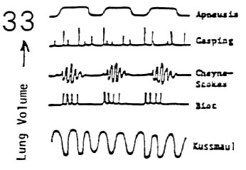
Kussmaul Breathing
Kussmaul breathing is a deep and labored breathing pattern often associated with severe metabolic acidosis, particularly diabetic ketoacidosis (DKA) but also renal failure. It is a form of hyperventilation, which is any breathing pattern that reduces carbon dioxide in the blood due to increased rate or depth of respiration.
Kussmaul breathing is respiratory compensation for a metabolic acidosis, most commonly occurring in diabetics in diabetic ketoacidosis. Blood gases on a patient with Kussmaul breathing will show a low partial pressure of CO2 in conjunction with low bicarbonate because of a forced increased respiration (blowing off the carbon dioxide). Base excess is severely negative. The patient feels an urge to breathe deeply, an "air hunger", and it appears almost involuntary.
A metabolic acidosis soon produces hyperventilation, but at first it will tend to be rapid and relatively shallow. Kussmaul breathing develops as the acidosis grows more severe. Indeed, Kussmaul originally identified this type of breathing as a sign of coma and imminent death in diabetic patients.
Duration of fasting, presence or absence of hepatomegaly and Kussmaul breathing provide clues to the differential diagnosis of hyperglycemia in the inborn errors of metabolism
Content courtesy: Wikipedia, Patterns of breathing: Loyola University Medical Education Network. Video from Youtube
Labels:
American Board of Pediatrics,
breathing patterns,
diabetes,
kussmaul breathing,
pediatric board exam,
pediatric endocrinology,
prakasam,
SackidMD
Thursday, February 16, 2012
Marfan Syndrome
Marfan syndrome an autosomal dominant genetic disorder of the connective tissue caused by mutations in the FBN1 gene on chromosome 15. People with Marfan's tend to be unusually tall, with long limbs and long, thin finger with dislocated lenses and aortic root dilation
FBN1, which encodes a connective protein called fibrillin-1
The Fibrillin 1 protein is essential for the proper formation of the extracellular matrix including the biogenesis and maintenance of elastic fibers. The extracellular matrix is critical for both the structural integrity of connective tissue but also serves as a reservoir for growth factors
In addition to being a connective protein that forms the structural support for tissues outside the cell, the normal fibrillin-1 protein binds to another protein, transforming growth factor beta (TGF-β)..TGF-β has deleterious effects on vascular smooth muscle development and the integrity of the extracellular matrix.Secondary to mutated fibrillin there is excessive TGF-β at the lungs, heart valves, and aorta, and this weakens the tissues and causes the features of Marfan syndrome.
Lens Dislocation:
In Marfan's the dislocation is typically superotemporal whereas in homocystinuria, the dislocation is inferonasal.
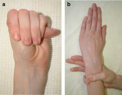
The Steinberg sign & Walker-Murdoch sign.
THE STEINBERG SIGN (a):
This test is used for the clinical evaluation of Marfan patients.
Procedure:
Instruct the patient to fold his thumb into the closed fist. This test is positive if the thumb tip extends from palm of hand (see figure a).
THE WALKER-MURDOCH SIGN (b):
This test is used for the evaluation of patients with Marfan syndrome.
Procedure:
Instruct the patient to grip his wrist with his opposite hand. If thumb and fifth finger of the hand overlap with each other, this represents a positive Walker-Murdoch sign (see figure b).
Courtesy: http://palmreadingperspectives.wordpress.com/2011/05/20/hands-signs-in-marfan-syndrome-thin-fingers-hand-shape-hypermobility/ and wikipedia
Labels:
American Board of Pediatrics,
fibrillin,
lens dislocation,
Marfan,
pediatric blog,
Pediatric Boards,
Pediatric cardiology,
prakasam,
SackidMD,
Steinberg Sign,
Walker-murdoch sign
Tuesday, February 14, 2012
Thyroid Antibodies
Thyroid peroxidase or thyroperoxidase (TPO) is an enzyme expressed mainly in the thyroid that liberates iodine for addition onto tyrosine residues on thyroglobulin for the production of thyroxine (T4) ortriiodothyronine (T3), thyroid hormones. In humans, thyroperoxidase is encoded by the TPO gene.
Thyroid peroxidase is a frequent epitope of autoantibodies in autoimmune thyroid disease, with such antibodies being called anti-thyroid peroxidase antibodies (anti-TPO antibodies).
| THYROID ANTIBODY | ACRONYM | PRESENT IN | WHEN ORDERED | OTHER FACTS |
|---|---|---|---|---|
| Thyroid peroxidase antibody | TPOAb | Hashimoto thyroiditis;Graves disease | When patient has symptoms suggesting hypothyroidism; when considering starting a patient on a drug therapy that has associated risks of developing hypothyroidism when thyroid peroxidase antibodies are present, such as lithium, amiodarone, interferon alpha, or interleukin-2 | Has been associated with reproductive difficulties, such as miscarriage,preeclampsia, premature delivery, and in-vitro fertilization failure |
| Thyroglobulin antibody | TgAb | Thyroid cancer; Hashimoto thyroiditis | Whenever a thyroglobulin test is performed to see if the antibody is present and likely to be interfering with the test results; since the thyroglobulin test will be ordered at regular intervals after thyroid cancer treatment, thyroglobulin antibody will also be ordered at regular intervals | |
| Thyroid stimulating hormone receptor antibody | TRAb | Graves disease | When patient has symptoms of hyperthyroidism; to monitor effectiveness of anti-thyroid therapy |
Labels:
American Board of Pediatrics,
pediatric board exam,
pediatric endocrinology,
prakasam,
SackidMD,
Thyroid antibodies
Monday, February 13, 2012
Wolff–Parkinson–White syndrome
Wolff-Parkinson-White syndrome is a heart condition in which there is an extra electrical pathway (circuit) in the heart. The condition can lead to episodes of rapid heart rate
Wolff-Parkinson-White is one of the most common causes of fast heart rate disorders in infants and children.
In people with Wolff-Parkinson-White syndrome, there is an extra, or accessory, pathway that may cause a very rapid heart rate. This is called supraventricular tachycardia.
ECG may show an abnormality called a "delta" wave. ( see the short PR interval)
Wolff-Parkinson-White is one of the most common causes of fast heart rate disorders in infants and children.
In people with Wolff-Parkinson-White syndrome, there is an extra, or accessory, pathway that may cause a very rapid heart rate. This is called supraventricular tachycardia.
ECG may show an abnormality called a "delta" wave. ( see the short PR interval)
Intracardiac electrophysiology study (EPS) : May be needed sometimes to make the diagnosis. The study involves placing wire electrodes in the heart to check for abnormal heartbeats or heart rhythms.
Medication may be used to control or prevent rapid heart beating. These include adenosine, antiarrhythmics, and amiodarone.
electricalcardioversion may be needed in some
The current preferred therapy for Wolff-Parkinson-White syndrome is catheter ablation (success rate for the procedure ranges between 85 - 95%). The small area that is causing the fast heart rate is destroyed using radiofrequency.
Rarely surgery is needed
Success rate of Catheter ablation will vary depending on location of accessory pathway and number of accessory pathways.
Courtesy: http://en.wikipedia.org/wiki/Wolff%E2%80%93Parkinson%E2%80%93White_syndrome, http://www.nlm.nih.gov/medlineplus/ency/article/000151.htm AND http://pedicardiology-bala.blogspot.com/2011/08/ekg-wpw-syndrome.html
Labels:
ABP,
Pediatric Boards,
Pediatric cardiology,
prakasam,
SackidMD,
Wolff-parkinson-White,
WPW
Sunday, February 12, 2012
Major Depressive Disorder in Children and Adolescents
Source: http://www.uspreventiveservicestaskforce.org/uspstf/uspschdepr.htm#summary
U.S. Preventive Services Task Force (USPSTF) recommendations on screening for depression in children and adolescents
Summary of Recommendations
- The USPSTF recommends screening of adolescents (12-18 years of age) for major depressive disorder (MDD) when systems are in place to ensure accurate diagnosis, psychotherapy (cognitive-behavioral or interpersonal), and follow-up.
- The USPSTF concludes that the current evidence is insufficient to assess the balance of benefits and harms of screening of children (7-11 years of age).
- Benefits of Detection and Early Intervention
- Adolescents (12-18 years of age): The USPSTF found adequate evidence that treatment in adolescents with selective serotonin reuptake inhibitors (SSRIs - fluoxetine) ), psychotherapy, and combined therapy (SSRIs and psychotherapy) results in decreases in MDD symptoms.
- Children (7-11 years of age): The USPSTF found inadequate evidence to support the benefits of treatment in children. SSRIs (fluoxetine) reduce MDD symptoms in children; however, there are limited data on the benefits of psychotherapy and the benefits of psychotherapy plus SSRIs in children.
- Assessment of Risk: Important risk factors that can be assessed relatively accurately and reliably include parental depression, having comorbid mental health or chronic medical conditions, and having experienced a major negative life event.
Screening Tests: Instruments developed for primary care (Patient Health Questionnaire for Adolescents [PHQ-A] and the Beck Depression Inventory-Primary Care Version [BDI-PC]) have been used successfully in adolescents.
There are limited data describing the accuracy of using MDD screening instruments in younger children (7-11 years of age).
Treatment: Among pharmacotherapies available for the treatment of MDD in children and adolescents, SSRIs have been found to be efficacious. Treating depressed youth with SSRIs is associated with an increased risk of suicidality and, therefore, should only be considered if judicious clinical monitoring is possible. Psychotherapy trials indicate that a variety of psychotherapy types are efficacious among adolescents (including cognitive-behavioral and interpersonal therapies). Harms of psychotherapy are felt to be small.
Update of Previous USPSTF Recommendation
This recommendation updates the previous recommendation released in 2002. The major change in the current recommendation is that the USPSTF now recommends screening of adolescents (12-18 years of age) for MDD when systems are in place to ensure accurate diagnosis, psychotherapy (eg, cognitive-behavioral, interpersonal), and follow-up. In 2002, the USPSTF concluded that there was insufficient evidence to recommend for or against routine screening of children or adolescents for MDD (I recommendation). The basis for this change in recommendation for adolescents is a result of new evidence that demonstrates treatment benefit.
Labels:
Depression,
fluoxetine,
Pediatric psychiatry,
prakasam,
SackidMD,
SSRI
Saturday, February 11, 2012
Glomerulonephritis (gloe-mer-u-lo-nuh-FRY-tis)
Glomerulonephritis, is a renal disease (usually of both kidneys) characterized by inflammation of the glomeruli, or small blood vessels in the kidneys.
It may present with isolated hematuria and/or proteinuria.
Presenting picture can be nephrotic syndrome, a nephritic syndrome, acute renal failure, or chronic renal failure.
Broadly grouped into non-proliferative or proliferative types.
Diagnosing the pattern of GN is important because the outcome and treatment differs in different types.
Primary causes are ones which are intrinsic to the kidney, whilst secondary causes are associated with certain infections (bacterial, viral or parasitic pathogens), drugs, systemic disorders (SLE, vasculitis) or diabetes.
Non Proliferative
This is characterised by the numbers of cells (lack of hypercellularity) in the glomeruli. They usually cause nephrotic syndrome. This has the following types:
Minimal change GN (also known as Minimal Change Disease)
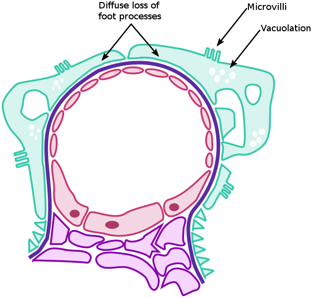
The three hallmarks of Minimal Change Disease: diffuse loss of podocyte foot processes, vacuolation, and the appearance of microvilli.
This form of GN causes >75% of nephrotic syndrome in children, but only ~20% in adults. There are no changes visible on simple light microscopy, but on electron microscopy there is fusion of podocytes (supportive cells in the glomerulus).
Immunohistochemistry staining is negative.
Treatment consists of supportive care for the massive edema and as well as steroids to halt the disease process (typically Prednisone 1 mg/kg). Over 90% of children respond well to steroids, being essentially cured after 3 months of treatment.
Adults have a lower response rate (80%).
Failure to respond to steroids ('steroid resistant') or return of the disease when steroids are stopped ('steroid dependent') may require cytotoxic therapy (such as cyclosporin) which is associated with many side-effects.
Other 2 typs with nonproliferative are Focal Segmental GN and Membranous glomerulonephritis.
Proliferative Glomerulonephritis
This type is characterised by increased number of cells in the glomerulus (hypercellular). Usually present as a nephritic syndrome and usually progress to end-stage renal failure (ESRF) over weeks to years (depending on type).
Courtesy: http://en.wikipedia.org/wiki/Minimal_change_disease and Mayo Clinic
It may present with isolated hematuria and/or proteinuria.
Presenting picture can be nephrotic syndrome, a nephritic syndrome, acute renal failure, or chronic renal failure.
Broadly grouped into non-proliferative or proliferative types.
Diagnosing the pattern of GN is important because the outcome and treatment differs in different types.
Primary causes are ones which are intrinsic to the kidney, whilst secondary causes are associated with certain infections (bacterial, viral or parasitic pathogens), drugs, systemic disorders (SLE, vasculitis) or diabetes.
Non Proliferative
This is characterised by the numbers of cells (lack of hypercellularity) in the glomeruli. They usually cause nephrotic syndrome. This has the following types:
Minimal change GN (also known as Minimal Change Disease)
The three hallmarks of Minimal Change Disease: diffuse loss of podocyte foot processes, vacuolation, and the appearance of microvilli.
This form of GN causes >75% of nephrotic syndrome in children, but only ~20% in adults. There are no changes visible on simple light microscopy, but on electron microscopy there is fusion of podocytes (supportive cells in the glomerulus).
Immunohistochemistry staining is negative.
Treatment consists of supportive care for the massive edema and as well as steroids to halt the disease process (typically Prednisone 1 mg/kg). Over 90% of children respond well to steroids, being essentially cured after 3 months of treatment.
Adults have a lower response rate (80%).
Failure to respond to steroids ('steroid resistant') or return of the disease when steroids are stopped ('steroid dependent') may require cytotoxic therapy (such as cyclosporin) which is associated with many side-effects.
Other 2 typs with nonproliferative are Focal Segmental GN and Membranous glomerulonephritis.
Proliferative Glomerulonephritis
This type is characterised by increased number of cells in the glomerulus (hypercellular). Usually present as a nephritic syndrome and usually progress to end-stage renal failure (ESRF) over weeks to years (depending on type).
Courtesy: http://en.wikipedia.org/wiki/Minimal_change_disease and Mayo Clinic
Labels:
glomerular nephritis,
minimal change,
nephrotic syndrome,
pediatric board exam,
Pediatric nephrology,
prakasam,
SackidMD
Friday, February 10, 2012
Interpretation of Hepatitis B Serologic Test Results
Hepatitis B serologic testing involves measurement of several hepatitis B
virus (HBV)-specifi c antigens and antibodies. Different serologic “markers”
or combinations of markers are used to identify different phases of HBV
infection and to determine whether a patient has acute or chronic HBV
infection, is immune to HBV as a result of prior infection or vaccination, or
is susceptible to infection.
Hepatitis B surface antigen (HBsAg):
A protein on the surface of hepatitis B virus; it can be detected in high levels in serum during acute or chronic hepatitis B virus infection. The presence of HBsAg indicates that the person is infectious. The body normally produces antibodies to HBsAg as part of the normal immune response to infection. HBsAg is the antigen used to make hepatitis B vaccine.
Hepatitis B surface antibody (anti-HBs):
The presence of anti-HBs is generally interpreted as indicating recovery and immunity from hepatitis B virus infection. Anti-HBs also develops in a person who has been successfully vaccinated against hepatitis B
Total hepatitis B core antibody (anti-HBc):
Appears at the onset of symptoms in acute hepatitis B and persists for life. The presence of
anti-HBc indicates previous or ongoing infection with hepatitis B virus in an undefined time frame
IgM antibody to hepatitis B core antigen (IgM anti-HBc):
Positivity indicates recent infection with hepatitis B virus (<6 mos). Its presence indicates acute infection.
Tests Results Interpretation
Susceptible
HBsAg negative
anti-HBc negative
anti-HBs negative
Immune due to natural infection
HBsAg negative
anti-HBc positive
anti-HBs positive
Immune due to hepatitis B vaccination
HBsAg negative
anti-HBc negative
anti-HBs positive
Acutely infected
HBsAg positive
anti-HBc positive
IgM anti-HBc positive
anti-HBs negative
Chronically infected
HBsAg positive
anti-HBc positive
IgM anti-HBc negative
anti-HBs negative
Interpretation unclear; four possibilities:
1. Resolved infection (most common)
2. False-positive anti-HBc, thus susceptible
3. “Low level” chronic infection
4. Resolving acute infection
anti-HBs negative
HBsAg negative
anti-HBc positive
Adapted from: A Comprehensive Immunization Strategy to Eliminate Transmission of Hepatitis B
Virus Infection in the United States: Recommendations of the Advisory Committee on Immunization
Practices. Part I: Immunization of Infants, Children, and Adolescents. MMWR 2005;54(No. RR-16
Courtesy: http://www.cdc.gov/hepatitis/HBV/PDFs/SerologicChartv8.pdf
virus (HBV)-specifi c antigens and antibodies. Different serologic “markers”
or combinations of markers are used to identify different phases of HBV
infection and to determine whether a patient has acute or chronic HBV
infection, is immune to HBV as a result of prior infection or vaccination, or
is susceptible to infection.
Hepatitis B surface antigen (HBsAg):
A protein on the surface of hepatitis B virus; it can be detected in high levels in serum during acute or chronic hepatitis B virus infection. The presence of HBsAg indicates that the person is infectious. The body normally produces antibodies to HBsAg as part of the normal immune response to infection. HBsAg is the antigen used to make hepatitis B vaccine.
Hepatitis B surface antibody (anti-HBs):
The presence of anti-HBs is generally interpreted as indicating recovery and immunity from hepatitis B virus infection. Anti-HBs also develops in a person who has been successfully vaccinated against hepatitis B
Total hepatitis B core antibody (anti-HBc):
Appears at the onset of symptoms in acute hepatitis B and persists for life. The presence of
anti-HBc indicates previous or ongoing infection with hepatitis B virus in an undefined time frame
IgM antibody to hepatitis B core antigen (IgM anti-HBc):
Positivity indicates recent infection with hepatitis B virus (<6 mos). Its presence indicates acute infection.
Tests Results Interpretation
Susceptible
HBsAg negative
anti-HBc negative
anti-HBs negative
Immune due to natural infection
HBsAg negative
anti-HBc positive
anti-HBs positive
Immune due to hepatitis B vaccination
HBsAg negative
anti-HBc negative
anti-HBs positive
Acutely infected
HBsAg positive
anti-HBc positive
IgM anti-HBc positive
anti-HBs negative
Chronically infected
HBsAg positive
anti-HBc positive
IgM anti-HBc negative
anti-HBs negative
Interpretation unclear; four possibilities:
1. Resolved infection (most common)
2. False-positive anti-HBc, thus susceptible
3. “Low level” chronic infection
4. Resolving acute infection
anti-HBs negative
HBsAg negative
anti-HBc positive
Adapted from: A Comprehensive Immunization Strategy to Eliminate Transmission of Hepatitis B
Virus Infection in the United States: Recommendations of the Advisory Committee on Immunization
Practices. Part I: Immunization of Infants, Children, and Adolescents. MMWR 2005;54(No. RR-16
Courtesy: http://www.cdc.gov/hepatitis/HBV/PDFs/SerologicChartv8.pdf
Thursday, February 9, 2012
Interesting Appendix!
One of the most common causes of severe acute abdominal pain worldwide.
A correctly diagnosed non-acute form of appendicitis is known as "rumbling appendicitis".
The term "pseudoappendicitis" is used to describe a condition mimicking appendicitis. It can be associated with Yersinia enterocolitica.
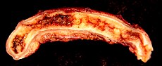
Picture shows an acutely inflamed and enlarged appendix, sliced lengthwise.
Rovsing's sign
Continuous deep palpation starting from the left iliac fossa upwards (counterclockwise along the colon) may cause pain in the right iliac fossa, by pushing bowel contents towards the ileocaecal valve and thus increasing pressure around the appendix.
Psoas sign or "Obraztsova's sign"
is right lower-quadrant pain that is produced with either the passive extension of the patient's right hip (patient lying on left side, with knee in flexion) or by the patient's active flexion of the right hip while supine. The pain elicited is due to inflammation of the peritoneum overlying the iliopsoas muscles and inflammation of the psoas muscles themselves. Straightening out the leg causes pain because it stretches these muscles, while flexing the hip activates the iliopsoas and therefore also causes pain.
Obturator sign
If an inflamed appendix is in contact with the obturator internus, spasm of the muscle can be demonstrated by flexing and internal rotation of the hip. This maneuver will cause pain in the hypogastrium.
Dunphy's sign
Increased pain in the right lower quadrant with coughing
Kocher's (Kosher's) sign
From the history given, the appearance of pain in the epigastric region or around the stomach at the beginning of disease with a subsequent shift to the right iliac region.
Differential diagnosis
In children
Gastroenteritis, mesenteric adenitis, Meckel's diverticulitis, intussusception, Henoch-Schönlein purpura, lobar pneumonia, urinary tract infection, new-onset Crohn's disease or ulcerative colitis, pancreatitis, abdominal trauma from child abuse, distal intestinal obstruction syndrome in children with cystic fibrosis, typhlitis in children with leukemia;
Courtesy: Wikipedia: http://en.wikipedia.org/wiki/Appendicitis
A correctly diagnosed non-acute form of appendicitis is known as "rumbling appendicitis".
The term "pseudoappendicitis" is used to describe a condition mimicking appendicitis. It can be associated with Yersinia enterocolitica.
Picture shows an acutely inflamed and enlarged appendix, sliced lengthwise.
Rovsing's sign
Continuous deep palpation starting from the left iliac fossa upwards (counterclockwise along the colon) may cause pain in the right iliac fossa, by pushing bowel contents towards the ileocaecal valve and thus increasing pressure around the appendix.
Psoas sign or "Obraztsova's sign"
is right lower-quadrant pain that is produced with either the passive extension of the patient's right hip (patient lying on left side, with knee in flexion) or by the patient's active flexion of the right hip while supine. The pain elicited is due to inflammation of the peritoneum overlying the iliopsoas muscles and inflammation of the psoas muscles themselves. Straightening out the leg causes pain because it stretches these muscles, while flexing the hip activates the iliopsoas and therefore also causes pain.
Obturator sign
If an inflamed appendix is in contact with the obturator internus, spasm of the muscle can be demonstrated by flexing and internal rotation of the hip. This maneuver will cause pain in the hypogastrium.
Dunphy's sign
Increased pain in the right lower quadrant with coughing
Kocher's (Kosher's) sign
From the history given, the appearance of pain in the epigastric region or around the stomach at the beginning of disease with a subsequent shift to the right iliac region.
Differential diagnosis
In children
Gastroenteritis, mesenteric adenitis, Meckel's diverticulitis, intussusception, Henoch-Schönlein purpura, lobar pneumonia, urinary tract infection, new-onset Crohn's disease or ulcerative colitis, pancreatitis, abdominal trauma from child abuse, distal intestinal obstruction syndrome in children with cystic fibrosis, typhlitis in children with leukemia;
Courtesy: Wikipedia: http://en.wikipedia.org/wiki/Appendicitis
Tuesday, February 7, 2012
Neonatal pustular melanosis or Transient Neonatal pustular melanosis
Other Terminologies:
1.Transient neonatal pustulosis
2.Transient Neonatal pustular melanosis
3.Lentigines neonatorum
It is a benign cutaneous condition that presents at birth with 1- to 3-mm flaccid, superficial fragile pustules, some of which may have already resolved in utero, leaving pigmented macules.
It has distinctive features characterized by vesicles, superficial pustules, and pigmented macules. The lesions of transient neonatal pustular melanosis are present at birth.
Commonly seen: On the chin, neck, forehead, chest, buttocks, back, and on the palms and soles
Self resolves in about 2 days. More common in dark skinned neonates.
Diagnosis: Usually Clinical. A Tzanck smear with a cellular stain (eg, Wright-Giemsa stain) or Gram stain of the contents of a pustule reveals a predominance of neutrophils and occasional eosinophils and cellular debris
Image Courtesy: DermAtlas
1.Transient neonatal pustulosis
2.Transient Neonatal pustular melanosis
3.Lentigines neonatorum
It is a benign cutaneous condition that presents at birth with 1- to 3-mm flaccid, superficial fragile pustules, some of which may have already resolved in utero, leaving pigmented macules.
It has distinctive features characterized by vesicles, superficial pustules, and pigmented macules. The lesions of transient neonatal pustular melanosis are present at birth.
Commonly seen: On the chin, neck, forehead, chest, buttocks, back, and on the palms and soles
Self resolves in about 2 days. More common in dark skinned neonates.
Diagnosis: Usually Clinical. A Tzanck smear with a cellular stain (eg, Wright-Giemsa stain) or Gram stain of the contents of a pustule reveals a predominance of neutrophils and occasional eosinophils and cellular debris
Image Courtesy: DermAtlas
Thursday, February 2, 2012
Diphtheria
Caused by Corynebacterium diphtheriae.
The bacteria most commonly infects the nose and throat. The throat infection causes a gray to black, tough, fiber-like covering, which can block the airways. In some cases, diphtheria may first infect the skin, producing skin lesions.
Signs and tests
The health care provider will perform a physical exam and look inside your mouth. This may reveal a gray to black covering (pseudomembrane) in the throat, enlarged lymph glands, and swelling of the neck or larynx.
Tests used may include:
Gram stain or throat culture to identify Corynebacterium diphtheriae
Electrocardiogram (ECG)
Treatment
If the health care provider thinks you have diphtheria, treatment should be started immediately, even before test results are available.
Diphtheria antitoxin is given as a shot into a muscle or through an IV (intravenous line). The infection is then treated with antibiotics, such as penicillin and erythromycin.
The death rate is 10%
Complications
The most common complication is inflammation of the heart muscle (myocarditis). The nervous system is also frequently and severely affected, which may result in temporary paralysis.
The diphtheria toxin can also damage the kidneys.
Courtesy of Centers for Disease Control and Prevention AND NIH
Subscribe to:
Posts (Atom)