Showing posts with label ABP. Show all posts

Tuesday, March 13, 2012

History of HbA1c and what does it do?

History:
Hemoglobin A1c was first separated from other forms of hemoglobin by Huisman and Meyering in 1958.  It was first characterized as a glycoprotein by Bookchin and Gallop in 1968. Its increase in diabetes was first described in 1969 by Samuel Rahbar The use of hemoglobin A1c for monitoring the degree of control of glucose metabolism in diabetic patients was proposed in 1976 by Anthony Cerami, Ronald Koenig and coworkers.
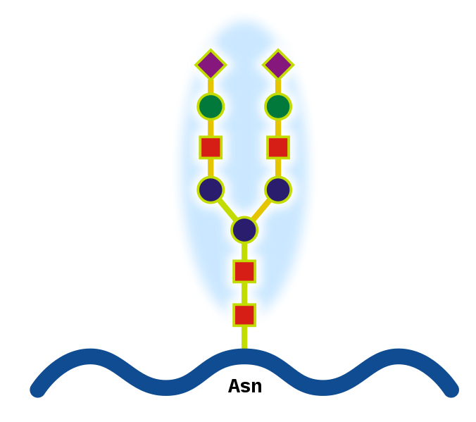
N-linked protein glycosylation (N-glycosylation of N-glycans) at Asn residues (Asn-x-Ser/Thr motifs) in glycoproteins.


What are Glycoproteins? 
They are proteins that contain oligosaccharide chains (glycans) covalently attached to polypeptide side-chains. The carbohydrate is attached to the protein in a cotranslational orposttranslational modification. This process is known as glycosylation. In proteins that have segments extending extracellularly, the extracellular segments are often glycosylated. Glycoproteins are often important integral membrane proteins, where they play a role in cell–cell interactions. Glycoproteins are also formed in the cytosol, but their functions and the pathways producing these modifications in this compartment are less well understood


Why do we measure HbA1c every 3 months?

In the normal 120-day lifespan of the red blood cell, glucose molecules react with hemoglobin, forming glycosolated hemoglobin. In individuals with poorly controlled diabetes, the quantities of these glycosolated hemoglobins are much higher than in healthy people.
Once a hemoglobin molecule is glycosolated, it remains that way. A buildup of glycosolated hemoglobin within the red cell, therefore, reflects the average level of glucose to which the cell has been exposed during its life-cycle. Measuring glycosolated hemoglobin assesses the effectiveness of therapy by monitoring long-term serum glucose regulation. The HbA1c level is proportional to average blood glucose concentration over the previous four weeks to three months. Some researchers state that the major proportion of its value is related to a rather shorter period of two to four weeks.
Image source: http://adiscar-adiscar.blogspot.com

We will talk about measuring HbA1c tomorrow

Monday, March 12, 2012

Tummy Tuck and Stretch Marks!

Medical Term for Strech Marks: Striae
Stretch marks or striae (singular stria), are a form of scarring on the skin with an off-color hue. They are caused by tearing of the dermis, which over time may diminish, but will not disappear completely.
Stretch marks are often the result of the rapid stretching of the skin associated with rapid growth.
Stretch marks can appear anywhere on the body, but are most likely to appear in places where larger amounts of fat are stored. Most common places are the abdomen, breasts, upper arms, underarms, back, thighs , hips, and buttocks. They pose no health risk in and of themselves.


In CUSHING SYNDROME, the glucocorticoid hormones responsible for the development of stretch marks affect the dermis by preventing the fibroblasts from forming collagen and elastin fibers, necessary to keep rapidly growing skin taut. This creates a lack of supportive material, as the skin is stretched and leads to dermal and epidermal tearing.
A new modality, fractional laser resurfacing, offers a novel approach to treating striae. Using scattered pulses of light only a fraction of the scar is zapped by the laser over the course of several treatments. This creates microscopic wounds. The body responds to each treatment by producing new collagen and epithelium


Picture courtesy: Derm Atlas


Abdominoplasty or "tummy tuck" is a cosmetic surgery procedure used to make the abdomen more firm. The surgery involves the removal of excess skin and fat from the middle and lower abdomen in order to tighten the muscle and fascia of the abdominal wall. 

Thursday, March 8, 2012

Who am I and why should you be scared of me now?

I am a very common virus that leads to mild, cold-like symptoms in adults and older healthy children. But I am bad with young babies. Most infants have been infected by me before their second birthday.
I love the season from October to Spring (my season!)

More severe  disease with me may occur in the following infants:
  • Premature infants
  • Infants with chronic lung disease
  • Immunodeficient infants
  • Infants with heart disease


The following increase the risk for being affected by me:
  • Day care
  • Tobacco smoke
  • Having school-aged brothers or sisters
  • Crowded conditions

    You get to know about me 4 - 6 days after coming in contact with me.
    Antibiotics cannot make me go away
    If am nice (mild) I go away without treatment.
    Infants and children with a severe infection with me will be in the intensive care unit. Treatment will include:
    • Oxygen ( may need a ventilator)
    • humidified air
    • IV fluids
    I AM Respiratory Syncytial Virus
    Belonging to the Paramyxoviridae, I am virus in an enveloped, spherical, negative-strand RNA virus measuring 120-300 nm

    Diagnosis: You can find me by a direct immunofluorescence test for RSV antigen , and is reported to be 90% sensitive as compared to culture. RSV antigen may appear in the cytoplasm of cells within 8 hours of infection. Viral culture is performed in conjunction with the antigen test. The only acceptable specimen to detect me is a nasopharyngeal aspirate collected in viral transport media.
    Tomorrow I will tell you how to avoid me!!

Wednesday, March 7, 2012

Turner Syndrome: Aortic Coarctation - see the 3D view

Turner's Syndrome: Chromosomal disorder - Turner syndrome affects approximately 1 out of every 2,500 female live births worldwide. It embraces a broad spectrum of features, from major heart defects to minor cosmetic issues. The commenest heart condition is BICUSPID AORTIC VALVE! not Coarctation of Aorta
See this study below:
Pediatr Cardiol. 1999 Mar-Apr;20(2):108-12.: Study shows:
Partial anomalous pulmonary venous drainage (PAPVD; 2.9%), 
Aortic valve disease (stenosis and/or incompetence) (AoVD; 5. 1%), 
Aortic coarctation (AoCo; 4.4%), 
Bicuspid aortic valve (BicAo; 14.7%) are much more frequent in Turner's syndrome than in the normal population, with the difference being statistically highly significant.


High Blood pressure is common because of heart or kidney issues
See and learn below: Magnetic resonance angiography  showing the aortic coarctation.


MR Angiography (MRA) - courtesy -http://turners.nichd.nih.gov/angiography.html

Thursday, March 1, 2012

Paul-Bunnell test - The other name for MONOSPOT test

The mononucleosis spot test looks for two antibodies in the blood that indicate infection with the Epstein-Barr virus (EBV).
. It is used to diagnosis infectious mononucleosis, a disease caused by the Epstein-Barr virus (EBV). About 1 week after the onset of the disease, many patients develop heterophile antibodies. Antibodies reach peak levels in 2 - 5 weeks and may persist for up to 1 year. However, a small number of persons with mononucleosis may never develop such antibodies.
Monospot tests are usually positive in approximately 85% of patients with infectious mononucleosis. Positive test results will not occur until 1 - 2 weeks into the illness.


False-positive results may be occur in persons with:
  • Hepatitis
  • Leukemia or lymphoma
  • Rubella
  • Systemic lupus erythematosus (SLE)

Algorithm for the management of suspected infectious mononucleosis. (IM = infectious mononucleosis; GABHS = group A β-hemolytic streptococcus; VCA = viral capsid antigen; EBV = Epstein-Barr virus)
Courtesy: http://www.aafp.org/afp/2004/1001/p1279.html

Sunday, February 26, 2012

Necrotising enterocolitis - Radiological findings

Supine abdominal x-rays are the classical picture needed for diagnosis. 
If NEC is suspected clinically, or there is concern on supine films the following can be done:
-cross-table lateral  or 
-left-lateral decubitus film


What do you see in the pictures?
1. Asymmetrically distributed dilated bowel loops
2.bowel wall edema with thumb printing
3.pneumatosis intestinalis (intramural gas)
4.portal venous gas
5.pneumoperitoneum
6. Free gas if there is peritoneal perforation


Source: Wiki Commons: RadsWiki

GOT SALT? Sodium Claims on Food Labels Claim - What they really mean?

Sodium Claims on Food Labels Claim
Explains what sodium claims on food labels such as “sodium-free” or “low-sodium” mean, as defined in federal guidelines.


“Sodium-free”:                                                  Less than 5 mg sodium per serving
“Salt-free”:                                                          Meets requirements for sodium-free
“Low-sodium”:                                                  140 mg sodium or less per serving
“Very low sodium”:                                          35 mg sodium or less per serving
“Reduced sodium”:                                          At least 25 percent less sodium when compared with the   regular version
“Light in sodium”:                                            50 percent less sodium per serving; restricted to foods with more than 40 calories per serving
“Unsalted, no added salt: not a 
sodium-free food”:                                           No salt is added during processing; the labeled product resembles or substitutes for another product that is normally processed with salt


Source: United States Food and Drug Administration (FDA) Food Labeling Guide. IX. Appendix A: Definitions of Nutrient Content Claims. FDA website.www.fda.gov/Food/GuidanceComplianceRegulatoryInformation/GuidanceDocuments/FoodLabelingNutrition/FoodLabelingGuide/ucm064911.htm. Revised April 2008. Accessed June 21, 2010.


As a rough guide, the maximum amount of salt for babies and children is: (source: national health service, UK
  • less than 1g (0.4g sodium) a day from 0-12 months,
  • 2g (0.8g sodium) a day from 1-3 years,
  • 3g (1.2g sodium) a day from 4-6 years,
  • 5g (2g sodium) a day from 7-10 years, and
  • 6g (2.4g sodium) a day from 11 years.


Source: CDC

Current dietary guidelines recommend that adults in general should consume no more than 2,300 mg of sodium per day. However, if you are in the following population groups, you should consume no more than 1,500 mg per day.
  • You are 40 years of age or older.
  • You are African American.
  • You have high blood pressure.

Tips for Reducing Sodium in Your Diet

 Buy fresh, plain frozen, or canned "with no salt added" vegetables.
 Use fresh poultry, fish, and lean meat, rather than canned or processed types.
 Use herbs, spices, and salt-free seasoning blends in cooking and at the table.
 Cook rice, pasta, and hot cereals without salt. Cut back on instant or flavored rice, pasta, and cereal mixes, which usually have added salt.
 Choose "convenience" foods that are lower in sodium. Cut back on frozen dinners, pizza, packaged mixes, canned soups or broths, and salad dressings — these often have a lot of sodium.
 Rinse canned foods, such as tuna, to remove some sodium.
 When available, buy low- or reduced-sodium, or no-salt-added versions of foods.
 Choose ready-to-eat breakfast cereals that are lower in sodium.


Source:  http://www.nhlbi.nih.gov/hbp/prevent/sodium/tips.htm

Saturday, February 25, 2012

Cri du chat syndrome

Information Courtesy: www.ncbi.nlm.nih.gov
Photo: Wikipedia


Chromosome 5p deletion syndrome; 5p minus syndrome; Cat cry syndrome
The syndrome’s name is based on the infant’s cry, which is high-pitched and sounds like a cat.

  • Cry that is high-pitched and sounds like a cat
  • Downward slant to the eyes
  • Low birth weight and slow growth
  • Low-set or abnormally shaped ears
  • Mental retardation (intellectual disability)
  • Partial webbing or fusing of fingers or toes
  • Single line in the palm of the hand
  • Skin tags just in front of the ear
  • Slow or incomplete development of motor skills
  • Small head (microcephaly)
  • Small jaw (micrognathia)
  • Wide-set eyes
    Intellectual disability (mental retardation) is common. Half of children with this syndrome learn enough verbal skills to communicate. The cat-like cry becomes less noticeable over time.

    Support Groups

    5p- Society -- www.fivepminus.org
    Facial features of a patient with Cri du Chat syndrome at age of 8 months (A), 2 years (B), 4 years (C) and 9 years (D)

Friday, February 24, 2012

Brudzinski’s sign


Even though Brudzinski  (a very famous Polish Physician)  described several signs in patients with meningitis, Brudzinski’s neck sign is most the most used test and is  referred to as Brudzinski’s sign.
How to elicit this sign?
With the patient supine, the examiner places one hand behind the patient’s head and places the other hand on the patient’s chest. The examiner then raises the patient’s head (with the hand behind the head)
while the hand on the chest restrains the patient and prevents the patient from rising. Flexion of the
patient’s lower extremities (hips and knees) constitutes a positive sign. Brudzinski’s neck sign is more sensitive than Kernig’s sign. 
How is it caused?

This sign is caused by the  motor nerve roots irritation which pass through inflamed meninges because the roots are brought under tension.
Image from Wikipedia: Meninges of the central nervous system: dura mater, arachnoid, and pia mater.

Cystic Hygroma

A cystic hygroma is a congenital multiloculated lymphatic lesion that can arise anywhere, but is classically found in the left posterior triangle of the neck.his is the most common form of lymphangioma
Cystic hygromas occur in approximately 1% of fetuses between weeks 9 and 16 of pregnancy
Cystic hygromas can occur as an isolated finding or in association with other birth defect. Because the risk for a chromosome abnormality approaches 50% when a cystic hygroma is found prenatally, amniocentesis or CVS (chorionic villus sampling) can be performed to diagnose these conditions.Cystic hygromas that develop in the third trimester (after thirty weeks gestation) or in the postnatal period are usually not associated with chromosome abnormalities. There is a chance of recurrence after surgical removal of the cystic hygroma.
A common association is Turner Syndrome. Severe form of this is HYDROPS.

Image Courtesy: DermAtlas

Video: Youtube

Wednesday, February 22, 2012

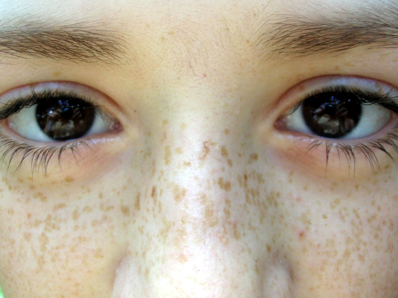
What the HECK is FRECKLE and how to differentiate fromLentigines

These are small pigmented spots on the skin with a clearly-defined edge, surrounded by normal-appearing skin. It is a harmless (benign) hyperplasia of melanocytes which is linear in its spread. is restricted to the cell layer directly above the basement membrane of the epidermis where melanocytes normally reside. 
This is in contrast to the "nests" of multi-layer melanocytes found in moles (melanocytic nevi). Because of this characteristic feature, the adjective "lentiginous" is used to describe other skin lesions that similarly proliferate linearly within the basal cell layer
The picture of lentigines below is from DERM ATLAS


Lentigines VS. Freckles:
Freckles have a relatively normal number of melanocytes but an increased amount of melanin. A lentigo has an increased number of melanocytes. Freckles will intensify and fade with sunlight exposure, whereas lentigines will stay stable in their color regardless of sunlight exposure
The pictures of Freckle below are from Wikipedia

Tuesday, February 21, 2012

Acanthosis nigricans

Darker, thick, velvety skin in body folds and creases. Rarely can cause itching


Commonest cause: Insulin Resistance (Insulin causes hyperplasia of the skin)
Internal Malignancies can cause this as well

Courtesy:

Avril Nimblett, MS, PA-C

 Acanthosis nigricans in children can be a precursor of type 2 diabetes


Image from Wikipedia

Monday, February 20, 2012

Anchovy sauce...

I know it is annoying to relate food to disease - but makes it easy remember and come with the correct diagnosis.
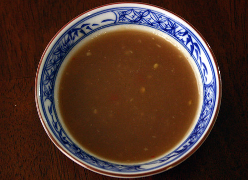
Anchovy sauce: Anchovies are a family of small, common salt-water  fish. Here you see Anchovy sauce in a bowl in the first picture. The picture underneath shows pus aspirated from a liver abscess confirmed to be due to entamoeba histolytica. 
Worldwide, Amoebic liver abscess is more common than pyogenic liver abscess.It is common in under developed countries. The disease is caused by ENTAMOEBA HISTOLYTICA. Infarction, enzymatic hydrolysis and immunological reaction either independently or in combination results in an abscess.The color has been described as “like anchovy sauce”. Anchovy sauce like pus is not always amoebic. It is not typical of amoebic liver abscess and has been over stressed in the past.The pus is usually chocolate colored or pinkish brown.
DIAGNOSTIC CRITERIA
1. History of residence in endemic area.
2. An enlarged tender liver in young male.
3. Response to metronidazole.
4. Leucocytosis
5. Elevation of right hemi diaphragm (radiology)
6. Scanning shows a filling defect.
7. A positive amoeba indirect haemagglutination test.

Source: AMOEBIC LIVER ABSCESS BY DR TAJUDDIN and wikipedia